
Blog
Minimally Invasive TLIF: What Doing a Fusion Through a Tube Really Means

A TLIF is a lumbar fusion that reaches the disc from the side, near the opening where the nerve leaves the spine. Minimally invasive means the muscle is spread apart with dilators rather than lifted off the bone, and the work is done through a tube about twenty millimeters wide with the screws placed through separate small openings under X-ray guidance.
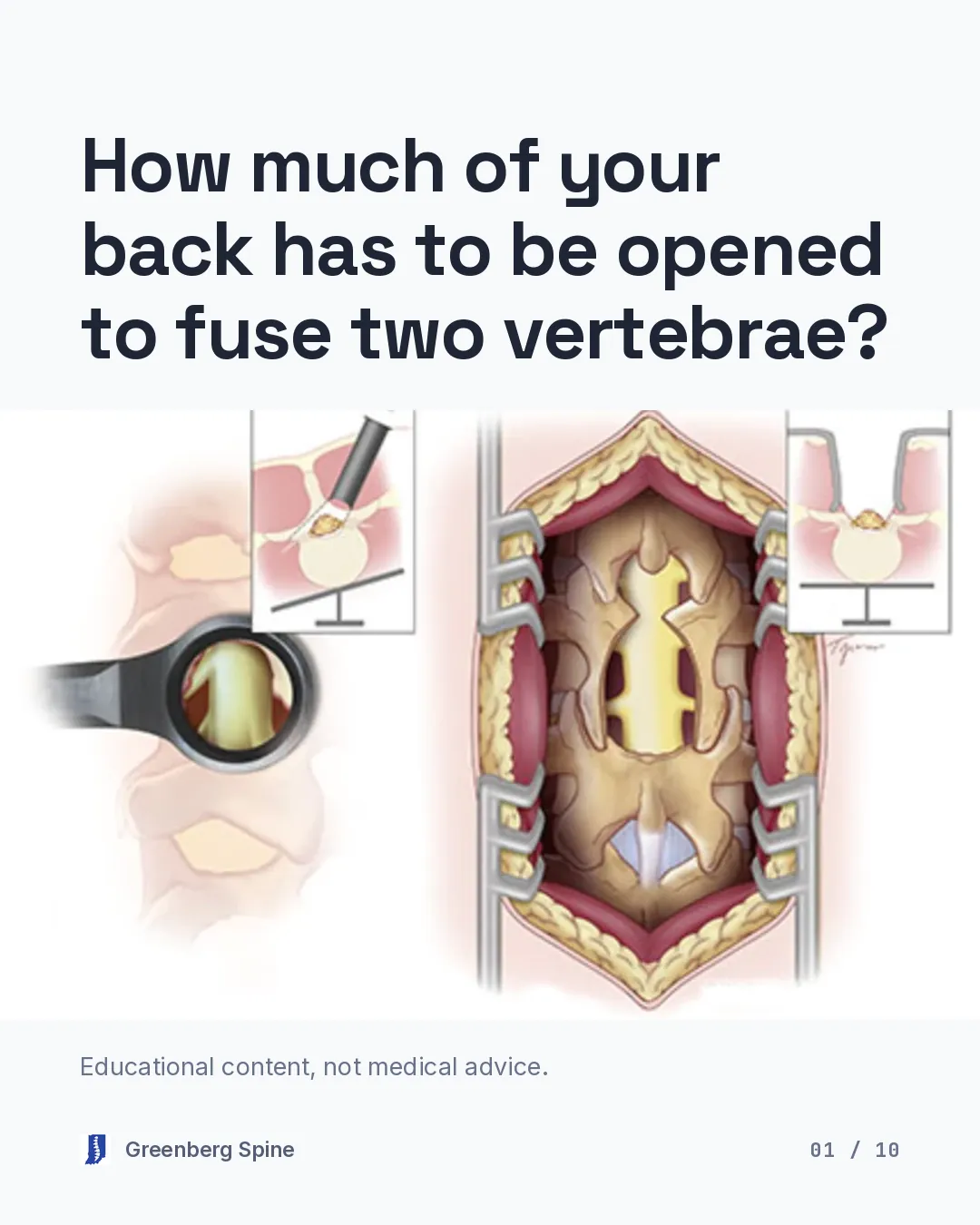
When someone is told they need a lumbar fusion, the question underneath the question is usually about the incision. How much of my back has to be opened to do this? It is a reasonable thing to want to know, and the answer has genuinely changed, though not in every way the phrase “minimally invasive” implies. So let me describe what actually happens, and then give you the fair version of how it compares with the open operation.
What a TLIF is
TLIF stands for transforaminal lumbar interbody fusion, and the first word is the one that carries the meaning. Transforaminal describes the route in: the disc is reached from the side, near the foramen, which is the opening where the nerve root leaves the spine. The worn disc comes out, a spacer called a cage goes into the space where it was, packed with bone graft, and screws and rods hold the two vertebrae still while they grow together into one segment. Our lumbar fusion page covers the operation in more detail, and if you are comparing routes into the spine, ALIF versus TLIF and lateral fusion versus TLIF take that up directly.
What “minimally invasive” actually means here
This is where the phrase gets slippery, so here is the concrete version. Minimally invasive describes how the surgeon gets to the spine, not what is done once there. The fusion itself is the same operation.
Instead of lifting the muscle off the bone to expose it, a series of dilators spreads the muscle apart, and a tube about twenty millimeters wide is docked onto the spine. Everything is done looking down that tube. The screws go in through separate small openings rather than through the same exposure, guided by X-ray images taken during the case. In a review of seventy-five studies describing this operation, 81 percent used a tube and 79 percent placed the screws that way, which is a reasonable definition of standard practice.
The planning is worth picturing too, because it explains the size of the incision. An X-ray taken in the operating room shows where the pedicles sit. Those are the strong bony tunnels the screws travel through. The incision is then marked one to two centimeters to the side of them, and it runs about three to four centimeters long.
Once inside, the surgeon removes the facet, the small joint at the back of the spine, on the side that hurts. That does two jobs at once: it takes the pressure off the nerve, and it opens a doorway into the disc for the cage.
Who it is considered for
A fusion is a bigger commitment than a decompression, so the reasons should be specific. The usual ones are a vertebra that has slipped out of line, which is spondylolisthesis, and a disc that has herniated again at a segment that has become unstable. A nerve pinched in the side opening can need that segment stabilized rather than just decompressed. So can a worn disc that still hurts after nonsurgical care has had a fair trial, which is one presentation of degenerative disc disease. Guidelines suggest adding a fusion when a slip comes together with narrowing that is causing symptoms.
The risks, stated plainly
Every version of this operation carries real risk, and going through a tube does not exempt it. In a review of 5,454 people who had the minimally invasive version, about 19 out of 100 had a complication of some kind. Nerve symptoms that later settled happened in about 2 out of 100, and lasting nerve injury in about 1 out of 100. The lining around the nerves can tear, and when it does it is repaired during the operation.
Two hardware problems deserve naming. Screws can miss the mark: about 96 out of 100 screws placed with X-ray guidance land within two millimeters of the plan, which means a small share do not, in a tunnel with a nerve root immediately beside it. And a cage can settle into the bone or shift out of position. If you are curious about what image guidance and robotics do and do not change here, we went through the evidence in robotic spinal fusion, myths versus evidence.
The longer-term risk is the segment next door. About 11 out of 100 people who have a lumbar fusion later develop symptoms at the level next to it. That is the standing cost of fusing a segment, and it belongs in the conversation before surgery rather than after.
Open surgery works, and the scores catch up
I want to be fair to the operation I am not describing, because a surgeon comparing techniques has an obvious thumb on the scale. Open surgery works. In a review of thirty-two studies, the differences that showed up were mostly about the operation itself and the days right after it: average blood loss was about 250 milliliters through the tube compared with about 570 with the open approach, and the average hospital stay was about two days shorter. At long-term follow-up, the pain and function scores came out the same.
There is also a trade-off in the other direction that gets left out of the brochure. Working through a tube means more X-ray time during the case, for you and for the operating team. And these techniques have a learning curve, which is a real variable in who should be doing them.
So the honest summary is that going through a tube changes the early part of the story more than the ending. That is worth something, and it is not everything. What recovery actually looks like week by week is covered in minimally invasive lumbar fusion recovery.
Before you agree to a fusion
Ask which of the numbers above apply to your spine specifically, and ask what the plan is if the fusion does not take. If a fusion has been recommended and you are not sure the reasoning has been explained to you, that is a good reason to get another set of eyes on the imaging.
Bring your MRI and your questions. I only operate when it’s truly the right thing for the patient, and when they’ve exhausted every other option first. You can request a consultation or ask for a second opinion, and told I need a spinal fusion is written for exactly that situation.
Sources
- TLIF: technique, indications and outcomes
- North American Spine Society guideline: degenerative lumbar spondylolisthesis
- Defining what is minimally invasive in lumbar interbody fusion
- MIS versus open TLIF: systematic review and meta-analysis
- MIS-TLIF technique and incision planning
- Complication rates of minimally invasive TLIF: review of the literature
- Fusion rates after MIS-TLIF and optimal graft material
- Accuracy of percutaneous pedicle screw placement
- Adjacent segment pathology after lumbar fusion
- MIS-TLIF learning curve and cage migration
- Fusion assessed at twelve months or later after interbody fusion
The slides, one at a time
These are the cards from the carousel version of this article. Each one stands on its own, so you can read them in order or stop at the one you came for.
Card 1 of 10 
Card 2 of 10 
Card 3 of 10 
Card 4 of 10 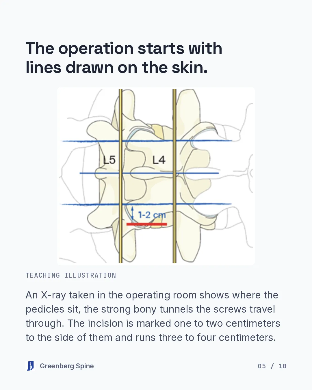
Card 5 of 10 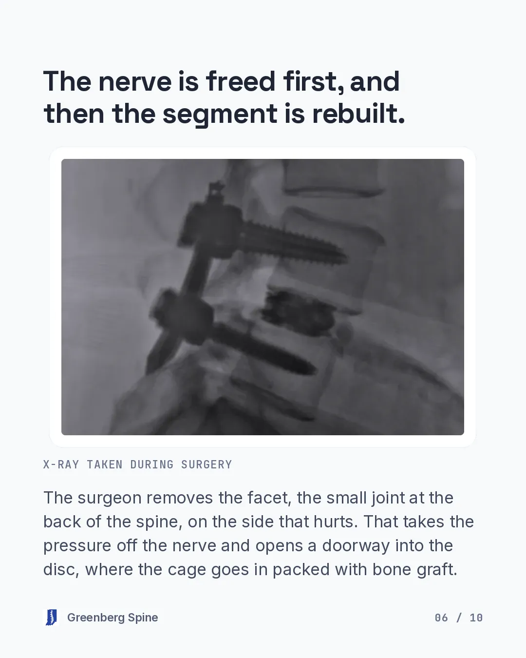
Card 6 of 10 
Card 7 of 10 
Card 8 of 10 
Card 9 of 10 
Card 10 of 10
This is general educational information, not medical advice. A clinical evaluation is the only way to know what’s right for you.